Home

çiftlik hayvanları Doğurmak zemin cholesteatoma diffusion mri amplifikatör cihaz yönetmek

Non-echoplanar diffusion weighted imaging in the detection of  post-operative middle ear cholesteatoma: navigating beyond the pitfalls to  find the pearl | Insights into Imaging | Full Text
Non-echoplanar diffusion weighted imaging in the detection of post-operative middle ear cholesteatoma: navigating beyond the pitfalls to find the pearl | Insights into Imaging | Full Text

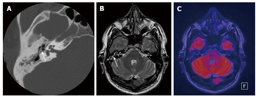
A case of left middle ear cholesteatoma. MRI shows hypointense on... |  Download Scientific Diagram
A case of left middle ear cholesteatoma. MRI shows hypointense on... | Download Scientific Diagram

EPOS™
EPOS™

Contemporary Non–Echo-planar Diffusion-weighted Imaging of Middle Ear  Cholesteatomas | RadioGraphics
Contemporary Non–Echo-planar Diffusion-weighted Imaging of Middle Ear Cholesteatomas | RadioGraphics

Cholesteatoma: multishot echo-planar vs non echo-planar diffusion-weighted  MRI for the prediction of middle ear and mastoid chol
Cholesteatoma: multishot echo-planar vs non echo-planar diffusion-weighted MRI for the prediction of middle ear and mastoid chol

Diffusion-Weighted Magnetic Resonance Imaging of Cholesteatoma Using  PROPELLER at 1.5T: A Single-Centre Retrospective Study - ScienceDirect
Diffusion-Weighted Magnetic Resonance Imaging of Cholesteatoma Using PROPELLER at 1.5T: A Single-Centre Retrospective Study - ScienceDirect

Contemporary Non–Echo-planar Diffusion-weighted Imaging of Middle Ear  Cholesteatomas | RadioGraphics
Contemporary Non–Echo-planar Diffusion-weighted Imaging of Middle Ear Cholesteatomas | RadioGraphics

Contemporary Non–Echo-planar Diffusion-weighted Imaging of Middle Ear  Cholesteatomas | RadioGraphics
Contemporary Non–Echo-planar Diffusion-weighted Imaging of Middle Ear Cholesteatomas | RadioGraphics
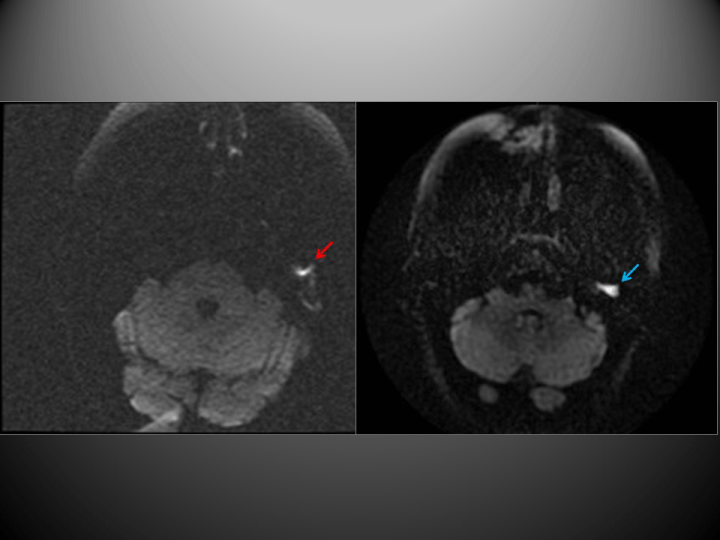
Diffusion-weighted magnetic resonance imaging with echo-planar and  non-echo-planar (PROPELLER) techniques in the clinical evaluation of  cholesteatoma
Diffusion-weighted magnetic resonance imaging with echo-planar and non-echo-planar (PROPELLER) techniques in the clinical evaluation of cholesteatoma

The value of different diffusion-weighted magnetic resonance techniques in  the diagnosis of middle ear cholesteatoma. Is there still an indication for  echo-planar diffusion-weighted imaging?
The value of different diffusion-weighted magnetic resonance techniques in the diagnosis of middle ear cholesteatoma. Is there still an indication for echo-planar diffusion-weighted imaging?
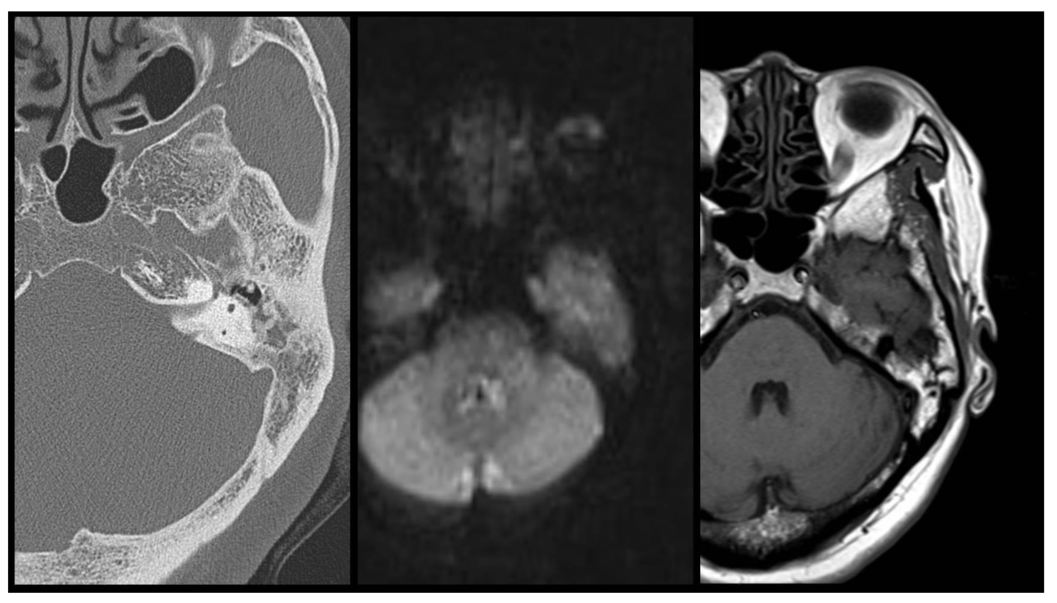
JPM | Free Full-Text | The Efficacy of DW and T1-W MRI Combined with CT in  the Preoperative Evaluation of Cholesteatoma
JPM | Free Full-Text | The Efficacy of DW and T1-W MRI Combined with CT in the Preoperative Evaluation of Cholesteatoma

BIR Publications
BIR Publications

Diffusion weighted imaging for the detection and evaluation of cholesteatoma

A 41-year-old male with a large cholesteatoma on the left side. MR... |  Download Scientific Diagram
A 41-year-old male with a large cholesteatoma on the left side. MR... | Download Scientific Diagram

Clinical Utility of MRI for Cholesteatoma Recurrence | SpringerLink

The Utility of Diffusion-Weighted Imaging for Cholesteatoma Evaluation |  American Journal of Neuroradiology
The Utility of Diffusion-Weighted Imaging for Cholesteatoma Evaluation | American Journal of Neuroradiology

Clinical Utility of MRI for Cholesteatoma Recurrence | SpringerLink

Role of diffusion-weighted MRI in the detection of cholesteatoma after  tympanoplasty - ScienceDirect
Role of diffusion-weighted MRI in the detection of cholesteatoma after tympanoplasty - ScienceDirect

Detection of Middle Ear Cholesteatoma by Diffusion-Weighted MR Imaging:  Multishot Echo-Planar Imaging Compared with Single-Shot Echo-Planar Imaging  | American Journal of Neuroradiology
Detection of Middle Ear Cholesteatoma by Diffusion-Weighted MR Imaging: Multishot Echo-Planar Imaging Compared with Single-Shot Echo-Planar Imaging | American Journal of Neuroradiology

Cholesteatoma of Temporal Bone - Stock Image - C027/1665 - Science Photo  Library
Cholesteatoma of Temporal Bone - Stock Image - C027/1665 - Science Photo Library

The Utility of Diffusion-Weighted Imaging for Cholesteatoma Evaluation |  American Journal of Neuroradiology
The Utility of Diffusion-Weighted Imaging for Cholesteatoma Evaluation | American Journal of Neuroradiology

Figure 1 from HASTE diffusion-weighted MRI for the reliable detection of  cholesteatoma. | Semantic Scholar
Figure 1 from HASTE diffusion-weighted MRI for the reliable detection of cholesteatoma. | Semantic Scholar

Role of diffusion-weighted MRI in the detection of cholesteatoma after  tympanoplasty - ScienceDirect
Role of diffusion-weighted MRI in the detection of cholesteatoma after tympanoplasty - ScienceDirect

The role of magnetic resonance imaging in the postoperative management of  cholesteatomas | Brazilian Journal of Otorhinolaryngology
The role of magnetic resonance imaging in the postoperative management of cholesteatomas | Brazilian Journal of Otorhinolaryngology

Cholesteatoma | Radiology Reference Article | Radiopaedia.org